
Developmental timeline
TS1 (fertilized egg) → P56 (adult) · click a stage to load samples
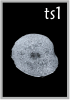
TS1
E0.5
1-cell
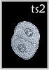
TS2
E1
2-cell
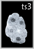
TS3
E2
4-16 cell
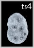
TS4
E3
morula → blastocyst
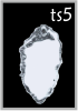
TS5
E4
late blastocyst
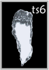
TS6
E4.5
attaching blastocyst
 1
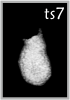
1TS7
E5
egg cylinder
 1
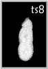
1TS8
E6
prestreak
 6
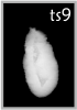
6TS9
E6.5
streak
 4
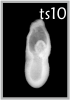
4TS10
E7
amnion
 6
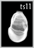
6TS11
E7.5
neural plate
 13
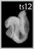
13TS12
E8
neural folds
 6
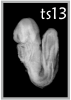
6TS13
E8.5
5-8 somites
 4
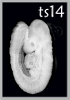
4TS14
E9
9-12 somites
 20
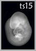
20TS15
E9.5
13-20 somites
 6
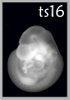
6TS16
E10
forelimb buds
 11
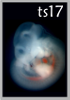
11TS17
E10.5
hindlimb buds
 9
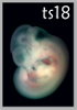
9TS18
E11
handplate
 17
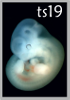
17TS19
E11.5
hindlimb handplate
 11
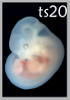
11TS20
E12
digital rays
 10
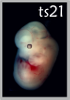
10TS21
E13.5
finger separation
 9
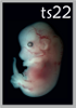
9TS22
E14
eyelid formation
 20
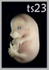
20TS23
E15
eyelids closing
 15
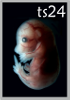
15TS24
E15.5
ear pinna
 26
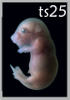
26TS25
E16.5
long whiskers
 2
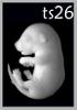
2TS26
E17.5
body hair
 1
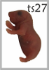
1TS27
E18.5
pre-birth
P7
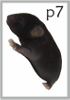
P7
postnatal day 7
P56
P56
adult (8 weeks)
E0
E2
E4
E6
E8
E10
E12
E14
E16
E18
Birth
E0E5E10E15BirthP7P56
TS11
E7.5· Neural plateAnterior neural plate appears. Heart field and somitic mesoderm specify.
2samples
1/2anatomy delineated
histology · 2Histology — embryo physically embedded and microtomed into 1–7 µm sections, stained (Nissl / H&E), imaged, and reassembled into a 3D volume. High in-plane resolution, anisotropic voxels. Stereo-seq · 2Stereo-seq (BGI) — DNB-array spatial transcriptomics with subcellular ~500 nm resolution. Captures whole-organ to whole-embryo sections; in the Spateo Cell 2024 atlas, 74 sections per embryo were aligned into a 3D cell-bin volume. imputed · 2Imputed — not measured. scRNA-seq cells placed into the eMouseAtlas anatomical mesh for this Theiler stage by canonical organ mapping. The cell-type-to-organ assignment is the imputed part; position within an organ is random, so organ-level structure is meaningful and sub-organ texture is not.
Transcriptomics data · 4
Stage-matched · not registered to atlas anatomy Stereo-seq
digiembryo_e7_5_rep1
13,168 cells · 17,172 genes
Spatial · mouse · stage TS11
Xie et al., Cell 2025 (digital embryo)
Open viewer →
Stereo-seq
digiembryo_e7_5_rep2
8,190 cells · 16,364 genes
Spatial · mouse · stage TS11
Xie et al., Cell 2025 (digital embryo)
Open viewer →
imputed
Imputed · E7.25
13,537 cells · 39 genes
Spatial · mouse · stage TS11 · dated E7.25, staged TS11
Imputed — eMouseAtlas mesh + TOME scRNA-seq
Open viewer →
imputed
Imputed · E7.5
10,994 cells · 39 genes
Spatial · mouse · stage TS11
Imputed — eMouseAtlas mesh + TOME scRNA-seq
Open viewer →
3D reference models
Histology plates · 50
Kaufman atlas, EMAPA-annotated · scroll → 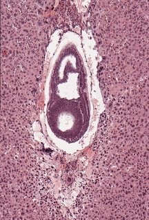 Sagittal12 ann
 Plate 05a
TS11
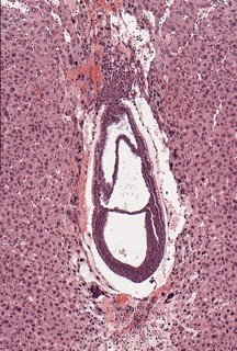 Sagittal4 ann
 Plate 05b
TS11
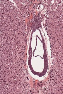 Sagittal6 ann
 Plate 05c
TS11
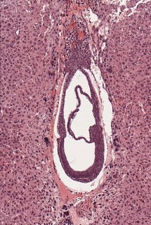 Sagittal5 ann
 Plate 05d
TS11
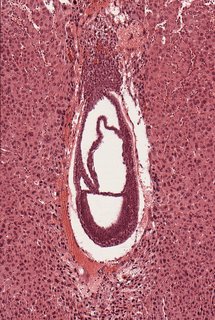 Sagittal3 ann
 Plate 05e
TS11
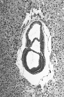 Sagittal3 ann
 Plate 05f
TS11
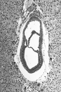 Sagittal5 ann
 Plate 05g
TS11
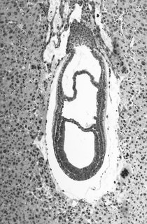 Sagittal4 ann
 Plate 05h
TS11
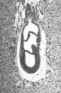 Sagittal6 ann
 Plate 05i
TS11
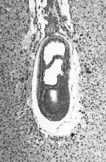 Sagittal4 ann
 Plate 05j
TS11
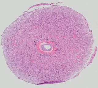 Transverse2 ann
 Plate 06a
TS11
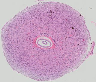 Transverse6 ann
 Plate 06b
TS11
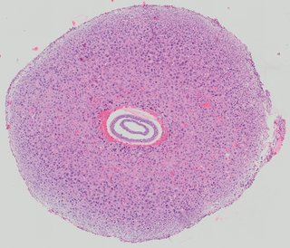 Transverse1 ann
 Plate 06c
TS11
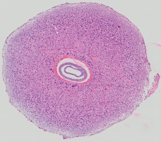 Transverse2 ann
 Plate 06d
TS11
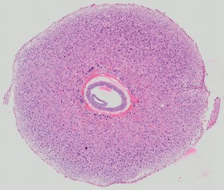 Transverse2 ann
 Plate 06e
TS11
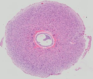 Transverse2 ann
 Plate 06f
TS11
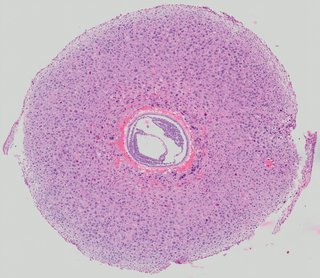 Transverse6 ann
 Plate 06g
TS11
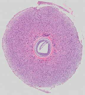 Transverse4 ann
 Plate 06h
TS11
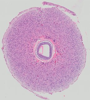 Transverse2 ann
 Plate 06i
TS11
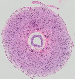 Transverse3 ann
 Plate 06j
TS11
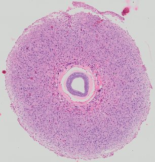 Transverse3 ann
 Plate 06k
TS11
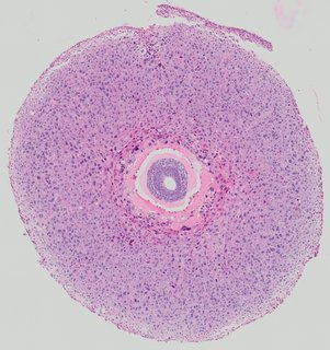 Transverse3 ann
 Plate 06l
TS11
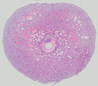 Transverse8 ann
 Plate 07a
TS11
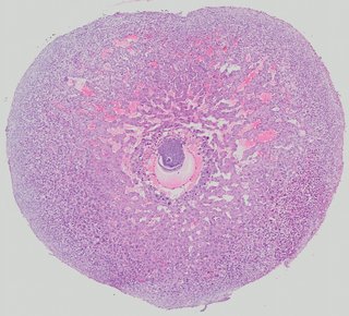 Transverse3 ann
 Plate 07b
TS11
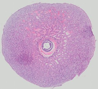 Transverse3 ann
 Plate 07c
TS11
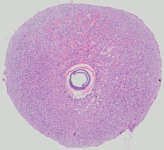 Transverse3 ann
 Plate 07d
TS11
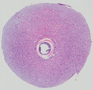 Transverse2 ann
 Plate 07e
TS11
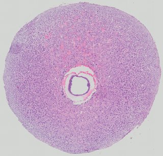 Transverse1 ann
 Plate 07f
TS11
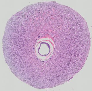 Transverse3 ann
 Plate 07g
TS11
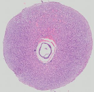 Transverse1 ann
 Plate 07h
TS11
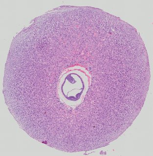 Transverse7 ann
 Plate 07i
TS11
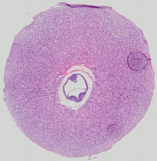 Transverse1 ann
 Plate 07j
TS11
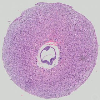 Transverse3 ann
 Plate 07k
TS11
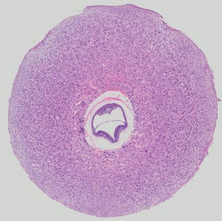 Transverse5 ann
 Plate 07l
TS11
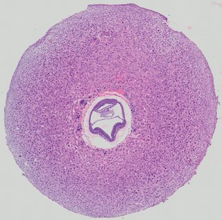 Transverse3 ann
 Plate 07m
TS11
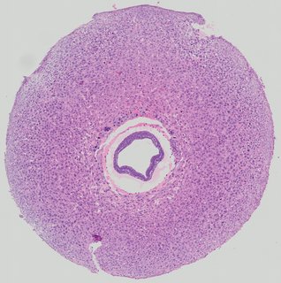 Transverse4 ann
 Plate 07n
TS11
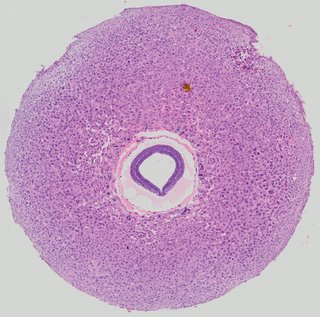 Transverse3 ann
 Plate 07o
TS11
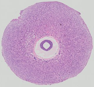 Transverse3 ann
 Plate 07p
TS11
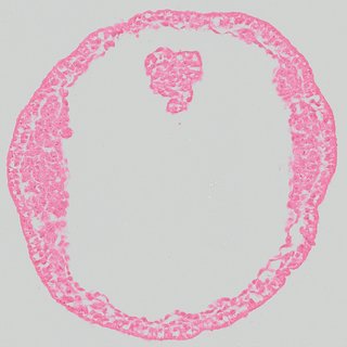 Transverse2 ann
 Plate 08a
TS11
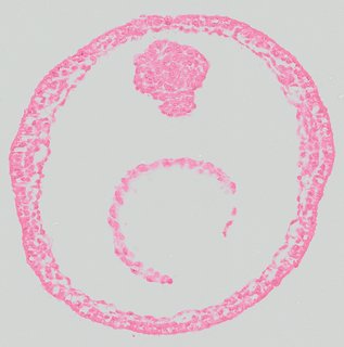 Transverse2 ann
 Plate 08b
TS11
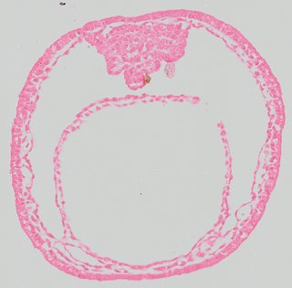 Transverse7 ann
 Plate 08c
TS11
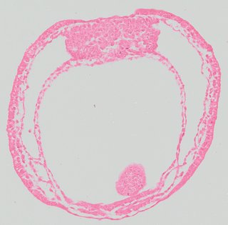 Transverse11 ann
 Plate 08d
TS11
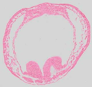 Transverse4 ann
 Plate 08e
TS11
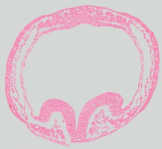 Transverse3 ann
 Plate 08f
TS11
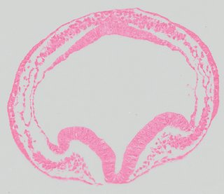 Transverse1 ann
 Plate 08g
TS11
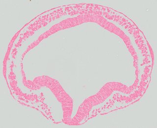 Transverse2 ann
 Plate 08h
TS11
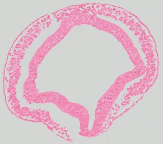 Transverse2 ann
 Plate 08i
TS11
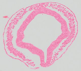 Transverse2 ann
 Plate 08j
TS11
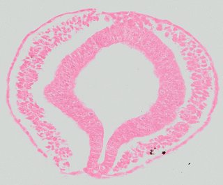 Transverse2 ann
 Plate 08k
TS11
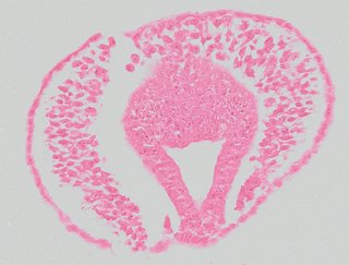 Transverse3 ann
 Plate 08l
TS11
Knowledge graph · TS11
EMAPA / CL-anchored · drag to rotate, scroll to zoom scRNA-seq · Literature · Predictions — coming soon
Previous
TS10E7
Amnion forms
About Theiler staging
Karl Theiler's staging system (1972) partitions prenatal development into 28 morphologically distinct steps. Virtual Embryo aligns all imagery, anatomy, and future spatial transcriptomics data to this axis.
Next
E8TS12
Neural folds
→